 Italiano
 English
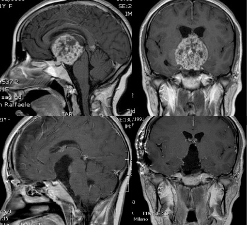
Craniopharyngioma is a type of brain tumor derived from pituitary gland embryonic tissue that occurs most commonly in children but also in men and women in their 50s and 60s. Craniopharyngiomas are relatively rare. Craniopharyngiomas can grow to large sizes, even bigger than a golf ball, and may not cause any symptoms in the beginning.
Symptoms
Craniopharyngiomas are typically very slow growing tumors. Symptoms produced by a craniopharyngioma vary depending upon the tumor’s location and the age of the patient. The symptoms also depend on which specific hormone is involved.
• Personality changes
• Headache
• Confusion
• Vomiting
• Pituitary insufficiency. This often occurs to some degree because craniopharyngiomas develop in the area of the pituitary stalk, which can affect the function of the pituitary gland.
In children, hormone deficiency can result in:
- Growth failure
- Delayed puberty
Other symptoms include:
- Diabetes insipidus. This occurs due to the absence of a posterior pituitary hormone called antidiuretic hormone (ADH).
Symptoms include:
- Excessive thirst
- Excessive urination
- Adrenal insufficiency. This occurs because of a reduction in ACTH production, a reduction in cortisol
In severe cases, it can be fatal. Symptoms include:
- Fatigue
- Low blood pressure
- Electrolyte abnormalities
• Growth hormone (GH) insufficiency, caused by the reduction in growth hormone (GH) production.
Symptoms include:
- Stunted growth and delayed puberty (in children)
- General fatigue, loss of muscle mass and tone (in adults)
• Hypothyroidism, caused by a reduction in TSH production. Symptoms include:
- Loss of appetite
- Weight gain
- Fatigue
- Decreased mental function
• Reduction in prolactin production. This is very uncommon and occurs with severe pituitary insufficiency.
- Large pituitary tumors can paradoxically elevate blood prolactin levels due to the “stalk effect.” This elevation occurs as a result of the compression of the pituitary stalk, which interferes with the brain’s control of prolactin production.
- In premenopausal women, elevated prolactin can lead to reduction or loss of menstrual periods and/or breast milk production (galactorrhea).
o With stalk effect, prolactin levels are usually only slightly elevated, as opposed to prolactinomas in which the prolactin level is usually very high. - Reduction of sex hormones, luteinizing hormone (LH) and follicle-stimulating hormone (FSH).
- In men, this can lead to a low testosterone level, causing decreased sexual drive and impotence.
- In some cases, there can be loss of body and facial hair.
- In women, this can lead to infertility
- If the tumor involves the optic tracts, chiasm, or nerves, blindness can occur.
If the hypothalamus, an area at the base of the brain, is affected, symptoms may include:
- Obesity
- Increased drowsiness
- Temperature regulation abnormalities
- Diabetes insipidus
Diagnosis
(MRI) scan the neuroradiologist to view the tumor from different angles. A CT scan can detect calcification in the tumor
Pituitary hormone function testing is necessary for every patient with a craniopharyngioma. An endocrinologist who specializes in pituitary tumors will help evaluate the results.
Treatment
Surgery
The two basic approaches are:
- Endonasal
- Craniotomy
Endoscopic Endonasal Surgery
The endoscopic endonasal approach is a minimally invasive approach, using your natural nasal passageway. It does not require a head incision. An endoscopic technique can be very effective in safely removing tumor, while at the same time minimizing hospitalization time and discomfort, however, not every craniopharyngioma tumor can be removed using an endonasal endoscopic approach.
Open Craniotomy
During an open craniotomy, the surgeon:
1. Makes an incision into your scalp
2. Temporarily removes a piece of your skull
3. Uses a surgical microscope to provide high magnification of the area
4. Access and remove the tumor; the most common approach involves gently elevating the base of the frontal lobe of the brain, above the eye.
A minimally invasive eyebrow incision keyhole approach can also be used.
Radiation Therapy for Craniopharyngiomas
Stereotactic radiosurgery is a technique that uses a highly focused dose of radiation delivered directly to the tumor. Because the radiation beam is sculpted to target only the tumor, the surrounding brain structures receive only a fraction of the radiation dose and may remain unharmed.
One drawback of radiation treatment is that it leads to delayed pituitary failure. This typically occurs several years after treatment, necessitating complete hormone replacement
Medical Therapy for Craniopharyngiomas
Pituitary hormonal dysfunction is common after craniopharyngioma surgery and/or radiation therapy. Problems with hypothalamic function can be particularly challenging to treat. You will require careful monitoring and follow-up visits with an endocrinologist.